| Paziente 1, con 8 Cd4/mmc. Pregressa pneumopatia intestiziale,
pericardite ed annessite. Isorgenza di febbre, cefalea, stabismo, diplopia, tosse. Mantoux
anamnesticamente positiva,. Iponatriemia. Reperti liquorali diagnostici per meningite
tubercolare, esame colturale positivo per Mycobacterium tuberculosis.
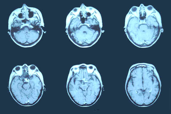
Fig. 1 - RMN. Presenza, in sede intra e sopra sellare, con estrinsecazione
parasellare sinistra, di tessuto a margini irregolarmente bozzoluti che mostra marcato e
disomogeneo enhancement dopo somministrazione di mdc. La lesione č responsabile di
compressione sul chiasma ottico e sul muso del terzo ventricolo. Presenza di aree nodulari
con marcato enhancement a carico delle cisterne della base, espressione di coinvolgimento
leptomeningeo. Deceduta dopo 2 mesi di terapia antitubercolare condotta in
maniera impropria per scarsa compliance.
Patient 1, with 8 Cd4/mmc. Previous interstitial pneumonia, pericarditis and adnexitis.
Onset of fever, headache, strabismus, diplopia, cough. History of positive Mantoux.
Hyponatraemia. Liquoral findings were diagnostic for tuberculous meningitis, cultural
examination was positive for Mycobacterium tuberculosis.
Fig. 1 - MRI. Presence, in intra and over sellar seat, with parasellar left
extension, of tissue with irregular margins, with marked inhomogeneous enhancement, and
with compression of optic chiasm and of the third ventricle. Presence of nodular areas
with marked enhancement of basal cisterns, expression of leptomeningeal involvement.
Dead after 2 months of inadequate antituberculous therapy for poor compliance. |