| Paziente 2. Cd4 300/mmc. Insorgenza di febbre e tosse. Esami
microbiologici negativi.
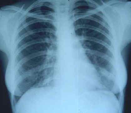
Fig. 1 - Rx. Opacitŕ disomogenea fusiforme, con margini sfumati, al terzo
inferiore del campo polmonare sinistro; interstiziopatia diffusa a vetro smerigliato;
minimo interessamento pleurico omolaterale.
Trattata con macrolidi e chinolonici con remissione clinica. Dopo 5 mesi ricomparsa di
febbre elevata, tosse ed emoftoe. Espettorato positivo per BK.
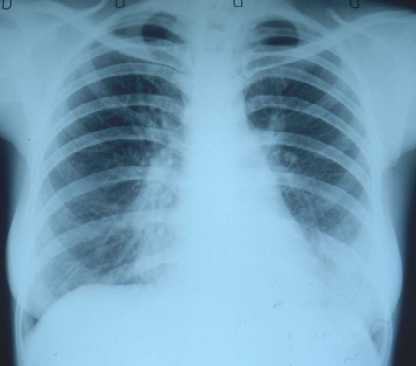
Fig. 2 - Rx. Opacitŕ disomogenea, con contorni sfumati alla base sinistra; ili
addensati ed ingranditi, interessamento interstizioalveolare diffuso, con aspetto
reticolonodulare, specialmente in sede parailare, bilateralmente.
Trattata con terapia antitubercolare classica, con remissione clinica.
Dopo 3 anni, insorgenza di febbre, tosse ed emoftoe.
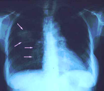
Fig. 3 - Rx. Opacitŕ parenchimali, con immagini escavative, al terzo medio del
campo polmonare destro.
Terapia antitubercolare classica praticata con successo clinico.
Patient 2. Cd4 300/mmc. Onset of fever and cough. Microbiological examinations were
negative.
Fig. 1 - X-ray film. Inhomogeneous fusiform opacity, with ill-defined margins, to
the lower third of left pulmonary field; widespread interstitiopathy with ground glass
pattern; mild homolateral pleural involvement.
Treated with macrolide and quinolonic with clinical remission. After 5 months onset of
fever of high degree, cough and haemoptysis. Sputum positive for BK.
Fig. 2 - X-ray film. Ill-defined inhomogeneous opacity, to the left pulmonary
base; enlarged hili, bilateral interstitio-alveolar involvement, with reticulonodular
pattern, specially in peri-hilar seat.
Treated with antituberculous therapy, with clinical remission.
After 3 years, onset of fever, cough and haemoptysis.
Fig. 3 - X-ray film. Parenchymal opacities, with cavitary images, to the middle
third of pulmonary right field.
Antituberculous therapy with clinical remission. |