| Paziente 1. 3 Cd4/mmc. Comparsa di febbre, tosse,
emiparesi destra e paralisi del settimo nervo cranico di sinistra. Lesioni cutanee
varicelliformi e zosteriformi. IgG toxo 260 UI. Esame dell'espettorato positivo per BK.
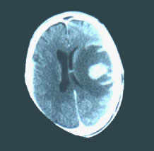
Fig. 1 - TC. Focolaio encefalitico con edema. Presenza in regione fronto-temporale
destra, di estesa area di ipodensitŕ con aree isodense e con effetto massa, con
compressione e spostamento delle strutture mediane: dopo mdc disomogeneo enhancement con
iperdensitŕ centrale di forma ovalare di circa 3 cm di diametro e di aspetto giriforme
alla periferia.
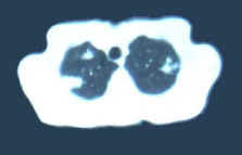
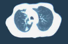
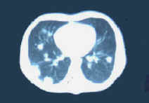
Fig. 2-4 -TC del torace. Presenza di multipli focolai di addensamento parenchimale
bilateralmente; adenopatia tracheobronchiale a destra.
Trattato con terapia antitoxoplasmica ed antitubercolare. Deceduto.
Patient 1, with 3 Cd4/mmc. Onset of fever, cough, right hemiparesis and paralysis of
the seventh nerve cranial of left. Vesicular and bullous cutaneous lesions for widespread
herpes zoster. IgG toxo 260 IU. Sputum examination positive for BK.
Fig. 1 - CT. Presence, in frontotemporal right region, of large area of
hypodensity with isodense areas with mass effect, compression and shift of middle
structures, inhomogeneous enhancement with central hyperdensities, oval in shape and 3 cm
of diameter, due to encephalitic focus with oedema.
Fig. 2-4 - CT. Presence of multiple foci of bilateral parenchymal consolidation;
right tracheobronchial adenopathy.
Treated with antitoxoplasma and antituberculous therapy. Dead. |